Quick Facts
- Survival Rate: The five-year relative survival rate is over 99% when the cancer is detected at a localized stage.
- Critical Threshold: Any spot with a diameter greater than 6mm, or roughly the size of a pencil eraser, warrants an immediate professional check.
- Risk Factor: Having 10 or more atypical moles can increase your personal risk of developing melanoma by 12 times compared to the general population.
- Age Warning: While moles are common in youth, any brand-new pigmented spots appearing after age 40 should be evaluated by a medical professional.
- Statistical Impact: Although melanoma accounts for only about 1% of skin cancers, it is responsible for the vast majority of skin cancer deaths.
- 2025 Projections: In the United States, an estimated 104,960 new cases of invasive melanoma are projected to be diagnosed in 2025.
- The Golden Rule: Evolution is the most significant clinical warning sign; any mole that changes in size, shape, or color is a priority for a dermatologist.
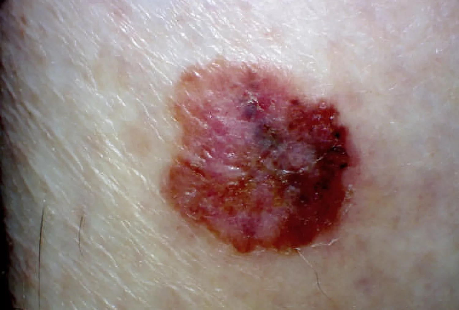
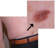
The ABCDE rule is a primary method for identifying melanoma warning signs. It includes checking for Asymmetry (one half doesn't match the other), Border irregularity (ragged or notched edges), Color variations (multiple shades or uneven distribution), Diameter (larger than 6mm), and Evolving (changes in size, shape, or color over time). Differentiating malignant melanoma from common sunspots or benign moles depends on consistency and change, making early detection screening a vital part of your preventive health routine.
The ABCDE Rule: Your Guide to Self-Detection
As a preventive care editor, I always tell my readers that the most powerful tool for skin health is your own eyes. While we often worry about external threats, the early signs of skin cancer are literally written on our skin. Understanding how to use the ABCDE rule for melanoma can transform a routine shower or dressing session into a life-saving self-exam. This mnemonic is designed to help you recognize the subtle shifts in pigmentation patterns that distinguish a healthy mole from a potential threat.
The first step is Asymmetry. If you were to draw an imaginary line through the center of a healthy mole, the two halves should look like twins. In melanoma, one half often looks entirely different from the other. Next is Border irregularity. Look for scalloped edges or margins that appear blurred, notched, or ragged. While a benign mole is usually a tidy circle or oval, the edges of a melanoma can look like a map of a rugged coastline.
Color is perhaps the most visually striking warning. A common mole is usually a single shade of brown. However, color variations are a major red flag. You might see different shades of brown, tan, or black, and as the lesion progresses, it may even show patches of red, white, or blue. Diameter is also key. We use the pencil eraser as our benchmark; anything larger than 6mm should be monitored closely. However, don't wait for a spot to grow if it meets other criteria, as many melanomas are caught when they are still small.
Finally, we look for Evolving. This is the most critical factor in preventive care. Signs of an evolving mole in melanoma include any change in size, shape, color, or elevation. If a spot starts to itch, crust, or bleed, it has moved beyond a simple cosmetic concern. When you perform a monthly skin self-check, you are looking for these shifts. I recommend using a full-length mirror and a hand mirror to check hard-to-reach areas like your back and the back of your thighs.
Melanoma vs. Sunspots: Spotting the Difference
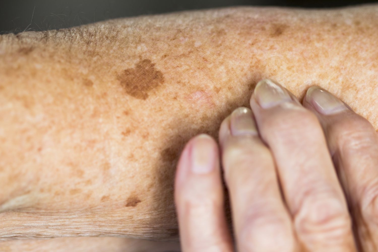
Many patients come to me confused by the brown spots that appear as we age. It is easy to mistake a benign lesion like a solar lentigo (common sunspot) for something more sinister. Sunspots are primarily a result of cumulative UV radiation risk over decades. They are typically uniform in color, flat, and remain stable for years. They are common on the face, hands, and shoulders—areas with high sun exposure.
The differences between melanoma and common sunspots become clear when you look at the life cycle of the spot. A sunspot arrives and stays mostly the same. A melanoma is dynamic. It is a biological "work in progress" that continues to alter its appearance. Furthermore, melanoma symptoms like itching or bleeding are rarely associated with sunspots. If a spot feels "angry" or persistently irritated without an obvious cause like friction from clothing, it needs a professional look.
| Feature | Sunspot (Solar Lentigo) | Malignant Melanoma |
|---|---|---|
| Symmetry | Usually symmetrical | Often asymmetrical |
| Border | Smooth and well-defined | Irregular, notched, or scalloped edges |
| Color | Uniform tan or brown | Multiple colors (black, red, blue, white) |
| Diameter | Varies, but remains stable | Often >6mm and grows |
| Texture | Flat and smooth | Can become raised or develop a crust |
| Sensations | None | Itching, tenderness, or bleeding |
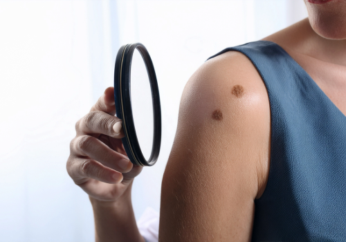
Atypical Moles and the 'Ugly Duckling' Sign
Not every concerning spot is a melanoma right away. Some are what we call dysplastic nevi, or atypical moles. These are unusual-looking benign moles that may resemble melanoma. People with many atypical moles have a higher risk of developing skin cancer, especially if they have a family history, a condition sometimes referred to as FAMMM syndrome. An atypical mole often has a fried egg appearance—a darker, raised center surrounded by a flatter, lighter tan area.
When looking for atypical mole symptoms, look for the "Ugly Duckling." The concept is simple: most moles on your body tend to look like each other. They are the same "family" of spots. The Ugly Duckling is the one mole that stands out because it is darker, larger, or a different shape than all the others. This outlier is often the one that requires a skin biopsy.
It is also vital to be aware of Amelanotic melanoma. This is a rare form of melanoma that lacks the dark pigment melanin. Instead of a dark brown or black spot, it might look like a pink, red, or skin-colored bump. Because it doesn't fit the traditional "dark mole" image, it is frequently overlooked. Atypical mole symptoms to watch for include any new, persistent growth that looks like a sore that won't heal or a shiny pink bump that wasn't there before.
Professional Diagnosis: When to See a Dermatologist
Self-exams are the first line of defense, but a Dermatologist is your partner in long-term wellness. If you find a spot that triggers any of the ABCDE criteria, schedule a professional evaluation immediately. You should also seek help for any lesion elevation that feels new or irregular, or if you notice any "bleeding" or "oozing" from a mole.
During your visit, the doctor may use a tool called a dermoscopy. This is a handheld device that provides a polarized, magnified view of the skin, allowing the doctor to see pigmentation patterns and structures invisible to the naked eye. If a lesion looks suspicious, the doctor will perform a skin biopsy. This involves removing a small sample of the tissue (or the entire lesion) to be examined under a microscope by a pathologist. This process, known as histopathology, is the only definitive way to confirm if a spot is malignant.
For those with a high number of moles, some clinics offer skin mapping. This involves high-resolution photography of your entire body to create a baseline. In future visits, the doctor can compare new photos to the old ones to see if any spots have moved or changed, which is an excellent way to catch an evolving mole before it becomes a major health issue.

FAQ
What are the early warning signs of melanoma?
The most common early signs include a change in an existing mole or the development of a new, unusual-looking growth. Using the ABCDE rule—looking for asymmetry, irregular borders, multiple colors, a diameter over 6mm, and any form of evolution or change—is the most effective way to identify these early indicators.
How can you tell the difference between a normal mole and melanoma?
Consistency is the hallmark of a normal mole; it usually stays the same size, shape, and color for years and matches other moles on your body. Melanoma is characterized by change and irregularity. If a mole stands out as different from the others (the Ugly Duckling) or shows rapid growth and uneven pigmentation, it is likely not a normal mole.
What are the ABCDEs of melanoma detection?
The ABCDEs are a mnemonic device: A for Asymmetry (mismatched halves), B for Border (ragged or scalloped edges), C for Color (multiple shades), D for Diameter (larger than 6mm), and E for Evolving (any change in the mole's appearance or symptoms like itching and bleeding).
Does a melanoma mole always have to be dark?
No, not all melanomas are dark. Amelanotic melanoma is a type that lacks pigment and can appear as a pink, red, or even skin-colored spot or bump. This is why any new or changing skin growth, regardless of its color, should be examined by a professional.
When should you see a dermatologist about a new spot?
You should see a dermatologist if a new spot appears after age 40, or if any spot at any age begins to change in size, shape, or color. Immediate consultation is required if a spot starts to itch, bleed, crust, or feel painful, as these can be signs of a more advanced lesion.
Protect Your Skin: Next Steps
Early detection is not just a medical recommendation; it is a lifestyle habit that ensures longevity and peace of mind. By integrating a monthly skin self-check into your routine, you become the expert on your own body. Remember to check areas often forgotten, such as the soles of your feet, between your toes, and your scalp.
Beyond self-checks, prioritize an annual professional screening. Think of it like a dental cleaning or an annual physical—it is a necessary checkpoint for your preventive care. Combine these screenings with sun-safe habits, like wearing broad-spectrum SPF and protective clothing, to minimize your UV radiation risk. Your skin is your body's largest organ and its first line of defense; giving it the attention it deserves is one of the best investments you can make in your long-term health.